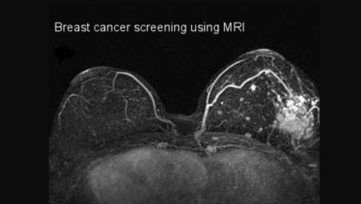
Breast cancer screening plays a crucial role in early detection and treatment. Radiology is at the heart of this process. It helps identify changes in breast tissue long before they become apparent. Using tools like mammograms and ultrasounds, radiologists detect potential issues with precision. Unlike knee pain Colorado , where the focus might be on joint imaging, breast cancer screening zeroes in on identifying tumors early. This early detection can save lives and improve outcomes.
Understanding Screening Techniques
Radiology uses various methods to screen for breast cancer. Let’s look at the three primary techniques:
- Mammograms: These are X-ray images of the breast. They can identify tumors that cannot be felt.
- Ultrasounds: These use sound waves to produce images and often help when mammograms show dense breast tissue.
- Magnetic Resonance Imaging (MRI): This technique uses magnets and radio waves. It provides detailed images and is usually reserved for high-risk cases.
These techniques can be combined for more comprehensive results. For example, some women may require both mammograms and ultrasounds, especially if they have dense breast tissue. The choice of technique often depends on individual risk factors and doctor recommendations.
The Role of Radiologists
Radiologists are the experts interpreting these images. They can spot subtle differences in breast tissue. Their expertise is crucial in identifying potential cancerous growths. This process involves careful comparison with previous images to detect any changes. According to the National Cancer Institute, regular mammograms reduce the risk of dying from breast cancer among women aged 40 to 74, especially those over 50.
Comparison of Screening Methods
| Method | Advantages | Disadvantages |
| Mammogram | Effective for early detection; widely available | Less effective for dense breasts; small radiation exposure |
| Ultrasound | No radiation; effective for dense breasts | Can miss small tumors; may require further testing |
| MRI | Highly detailed images; good for high-risk cases | Expensive; not typically used for screening |
Continuous Improvements
Advancements in technology continue to improve the accuracy of breast cancer screening. Digital mammography has replaced traditional film in many places. It allows radiologists to enhance images for better detail. Some centers now use 3D mammography, offering even clearer images.
Research is ongoing to refine these techniques further. For instance, the development of artificial intelligence tools offers promise. These tools can assist radiologists by highlighting areas of concern. As these technologies evolve, they may improve early detection and reduce false positives.
Importance of Regular Screenings
Regular screening remains a key factor in fighting breast cancer. Health organizations, such as the Centers for Disease Control and Prevention (CDC), recommend women between ages 50 and 74 have a mammogram every two years. Women with higher risk factors may need to start earlier or screen more often.
Early detection through screening offers the best chance of successful treatment. It can catch cancer before any symptoms arise, increasing the range of treatment options available.
Conclusion
Radiology plays a vital role in breast cancer screening. Effective imaging techniques, aid in early detection. This, in turn, improves treatment success rates and survival. As technology advances, radiology continues to evolve, offering hope for even better outcomes in the future.
